CHO cells
thingiverse
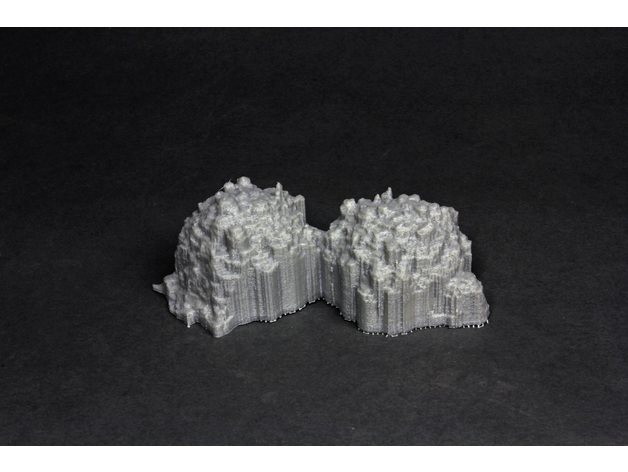
2 dividing wild-type Chinese hamster ovary (CHO) cells reconstructed from a scanning electron microscope data, 4000x magnification. The cell membrane on these is bubbly- packed with 'blebs'. Scanning electron microscope images by Jess Holz, 3D reconstruction by Ahmadreza Baghaie in the laboratory of Dr. Zeyun Yu, UW-Wisconsin Milwaukee Computer Science Department: https://pantherfile.uwm.edu/yuz/www/bmv/index.html How I Designed This 3D reconstruction from scanning electron microscope data Created by 3d reconstruction from a stereo-pair of scanning electron microscope images, taken at about 8 degrees tilt relative to each other. The algorithm creates a dense reconstruction in a manner not entirely unlike 123d catch. The algorithm was developed by the laboratory of Dr. Zeyun Yu, University of Wisconsin-Milwaukee Computer Science Department. See our latest paper by Tafti et al: 3DSEM++: Adaptive and intelligent 3D SEM surface reconstruction, available here: http://www.sciencedirect.com/science/article/pii/S0968432816300750 Original scanning electron microscopy data by Jess Holz 3d reconstruction.
With this file you will be able to print CHO cells with your 3D printer. Click on the button and save the file on your computer to work, edit or customize your design. You can also find more 3D designs for printers on CHO cells.