 NIH 3D
Coarctation, PAPVR
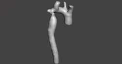
Demonstrated is a bicuspid aortic valve and history of coarctation repair within the first week of life by end to end anastomosis. MRI obtained for evaluation of distal arch.
MRI findings:
• PAPVR of left upper lobe to innominate vein: Qp:Qs of 1.4:1
• Mild residual narrowing of second transverse segment of the aortic arch.
• Moderate post-stenotic dilation of aorta
MRI images obtained at end-systole due to tachycardic heart rate during exam.
RV End-systolic volume is 36.3 ml.
LV End-systolic volume is 30.06 ml.
MRI methods:
A GE 1.5T HDxt system was used for the 3D HEART sequence which used a 3D respiratory-navigated balanced SSFP (steady state free precession) multi-slab sequence with T2 preparation that provides whole heart coverage with high contrast-to-noise ratio between vessels and myocardium. Due to the relatively fast heart rate of 122 bpm, the fat saturation was turned off to decrease the time needed for the prepatory pulse brining the acquisition window earlier into the cardiac cycle so that it could be centered on the quiescent stage of end systole. The sequence was run with the following parameters: TR 3.4, TE 1.4, Freq 224, Phase 160, RR 8, and fat sat off.
Learning:
The MRI identified previously un-diagnosed partial anomalous pulmonary venous return. However, the Qp:Qs fell within acceptable left to right shunting of < 1.5:1 and there was insignificant RV, RA enlargement. The MRI evaluation of the coarctation repair revealed a good repair with only mild narrowing, which appeared more severe by echo due to the post-stenotic dilation.
Disclaimer:
The available model has been validated to demonstrate the case’s pathologic features on a Z450 3D printer, (3DSystems, Circle Rock Hill, South Carolina)(or other printer as appropriate). While the mask applied to the original DICOM images accurately represents the anatomic features, some anatomic detail may be lost due to thin walled structures or inadequate supporting architecture; while other anatomic detail may be added due to similar limitations resulting in bleeding of modeling materials into small negative spaces. However, intracardiac structures, relationships, and pathologic features represent anatomic findings to scale and in high detail.
Direct link to the original creator's page
More from Matthew Bramlet


Similar Models
 NIH 3D
 NIH 3D
 NIH 3D
 Printables
 Thingiverse
 NIH 3D
 NIH 3D
 NIH 3D
Model Specifications
Frequently Asked Questions
Where is Coarctation, PAPVR published?
This model is published by its creator on NIH 3D. STLFinder only indexes it: the "View on NIH 3D" button above opens the original page, where the creator shares the STL, X3D, ZIP files under their own license.
What 3D printer can I use for this model?
This model is compatible with most FDM 3D printers (Creality Ender 3, Prusa MK3S+, Bambu Lab, etc.) and resin printers (Elegoo, Anycubic). Check the original page for recommended print settings and materials.
Is this 3D model free?
This is a premium model priced at 0 USD on NIH 3D. Premium models often include higher detail, multiple parts, and print-tested files.
Can I modify this file?
Check the license (CC-BY-NC-ND) on the original NIH 3D page. Most STL files can be modified using free software like Blender, TinkerCAD, or Meshmixer.