Dental Intra Oral X-ray Pos - question
sketchfab
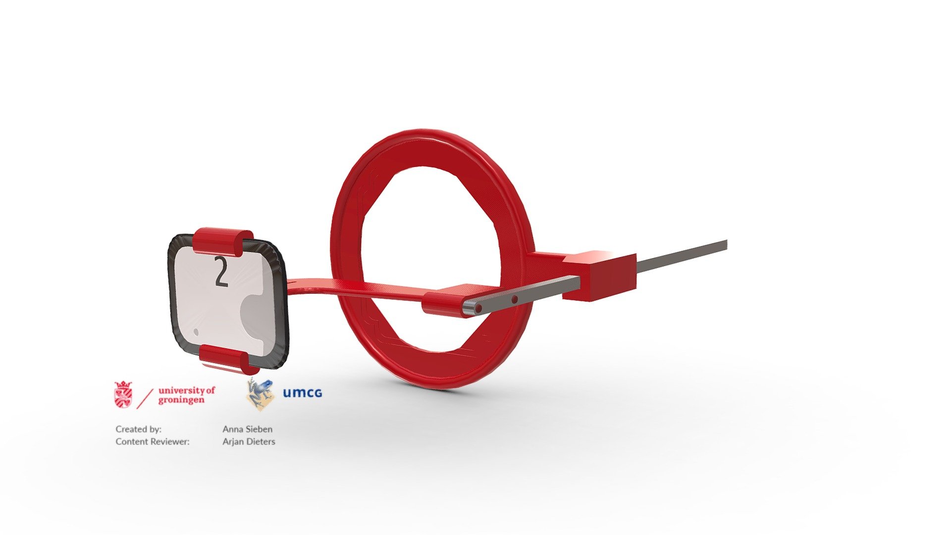
Dental X-rays (radiographs) are images of the teeth that a dentist uses to evaluate oral health. These X-rays are used with low levels of radiation to capture images of the interior of teeth and gums. This can help a dentist to identify problems, like cavities, tooth decay, and impacted teeth. This model depicts the positioning for a bite-wing x-ray. These x-rays show details of the upper and lower teeth in one area of the mouth. Each bite-wing shows a tooth from its crown (the exposed surface) to the level of the supporting bone. Bite-wing x-rays detect decay between teeth and changes in the thickness of bone caused by gum disease. Bite wing x-rays can also help determine the proper fit of a crown (a cap that completely encircles a tooth) or other restorations (eg, bridges). It can also see any wear or breakdown of dental fillings. This model is used in a digital handbook for a radiology practical for dentistry students.
With this file you will be able to print Dental Intra Oral X-ray Pos - question with your 3D printer. Click on the button and save the file on your computer to work, edit or customize your design. You can also find more 3D designs for printers on Dental Intra Oral X-ray Pos - question.