iPad Microscope Adapter
thingiverse
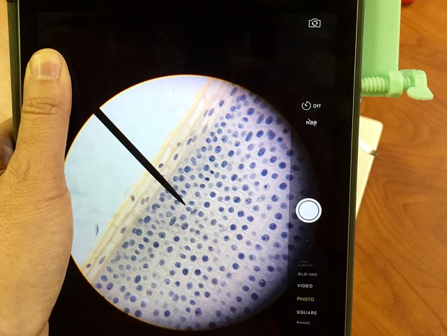
While walking around a biology class last year I noticed many students trying to use their iPads to take pictures through the eyepiece of their microscopes. While it is possible to get good images this way, it is incredibly difficult. This is my attempt to make it possible without as much luck needed. Being able to rest your iPad on a surface and then having two points to press against makes it much easier to keep the camera aligned looking through the eyepiece. Print Settings Printer Brand: MakerBot Printer: MakerBot Replicator Rafts: No Supports: No Resolution: 0.3 mm Infill: 10% Notes: I specifically didn't upload the models on a single build plate. I found the screws broke very easily while de-plating. I recommend printing them separate from the main piece with more perimeters and/or a higher infill. Conversely you can just be extra careful. You will obviously need to print two copies of the screw. Post-Printing No clean-up is needed. I originally designed an adapter for a naked iPad Air, but decided we now have several models of iPads in our school and most have cases. This design allows you to adjust for the particular iPad/case combination. Start by flipping the iPad so you can see the camera. Adjust the screws so that the camera is as close to the center of the hole as you can get it. Then mount the adapter to your microscope. You will need a little fine tuning and practice in order to capture some really nice images. The screws need to be adjusted slightly and/or you need to change move the iPad slightly so that your image is in a circle with a nice crisp edge to ensure the maximum field of view. Only a little practice is needed to get good images. How I Designed This I designed this in Tinkercad. It involved quite a bit of trial and error. You'll notice, for example, that the adapter keeps the iPad from getting too close to the eye piece. I learned that the vignetting gets worse the closer the camera lens is to the microscope. The screws are positioned such that they should not press any of the iPad's buttons while in use. I designed this with the iPad Air and the Air 2 in mind. So, I can't confirm this will not be a problem for the iPad Pro or older iPad models. If it is you can easily edit my model on Tinkercad. Our student microscopes have an eyepiece with a 28.5 mm diameter. In Tinkercad I used a 29 mm hole and it fits with a very nice level of tightness. If you change my design you'll still need to keep the iPad to be 10-15 mm away from the eyepiece for optimal viewing. The nut and bolt came from mike_linus' Nut Job. I imported the stl files into Tinkercad and mounted the nuts as seen and then added wings to the bolt to make it easier to turn. Although in retrospect, the wings aren't really needed. Tinkercad Models: Main Stand Screws Standards NGSS Overview and Background In a traditional biology class students often start learning about mitosis by looking at styalized/simplified pictures and they learn the names of the different stages. This leads to memorization without any real understanding. It would be much better to have students start by conducting investigations similar to the scientists who first described the process. In a very real sense, to “learn” the meaning of a new phrase or term requires that one “reinvent” or re-create the concept.” (Lawson, 1995) I propose having students look at well prepared specimens of onion root tips and have them look for patterns and differences. They should be able to see differences and sequence what they see. From this, the idea of cell division can be constructed in large part by the students themselves. The precise/accepted language can be introduced later. Objectives Students will begin to recognize the major stages in cell division Students will be able to put into the correct sequence a number of pictures showing a cell at various points undergoing mitosis. Audiences High school students Subjects Biology Skills/Standards NGSS - Disciplinary Core Ideas LS1.A: Structure and Function LS1.B: Growth and Development of Organisms NGSS - Scientific and Engineering Practices Asking Questions Developing and using models Analyzing and Interpreting Data NGSS Crosscutting Concepts Patterns Scale, proportion, and quantity Structure and Function Lesson Plan and Activity Have students in pairs photograph three to four areas on an onion root tip cross section at 400x magnification (using the 40x objective lens) In whole class discussion, ask students what they see? If possible have students share some of their pictures with the class with a projector or large computer screen. From this discussion you want to lead them to the realization that the different cells they see represent different points in a sequence. They should see changes in: Nucleus DNA Chromosomes With their partners, have students crop out individual cells that look different and try to sequence them. Apps that might be good for this: Notability Keynote Have pairs of students join each other (into groups of four) to share their pictures with each other. Ask such questions as: Are all of these different points in the sequence? What do you think is happening? Can you identify some “major” stages? Try to identify 4-6. Have students create sketches of their stages. They can also use an app like Adobe Ideas to trace the cell. Adobe Ideas uses layers, so the photo can be removed leaving just the sketch. Groups share results with the class. Draw student attention to the mitosis section in their textbooks and have them draw parallels between their sequence and the stages traditionally shown. Are all the traditional stages represented? Why not? Which if any are missing? Why? Duration 2 - 3 class periods Materials Needed Preparation Skills/Knowledge Required: Students should already be familiar with basic cell structure Students should know the basics of DNA and chromosomes Students should have a basic understanding of microscope use Stuff Needed: One microscope per pair of students One iPad microscope adapter and iPad per pair of students Prepared onion root tip slides stained to show mitosis References Lawson, A. E. (1995). Science teaching and the development of thinking. Belmont, CA: Wadsworth Pub.
With this file you will be able to print iPad Microscope Adapter with your 3D printer. Click on the button and save the file on your computer to work, edit or customize your design. You can also find more 3D designs for printers on iPad Microscope Adapter.