Photo 51: x-ray diffraction pattern of DNA
thingiverse
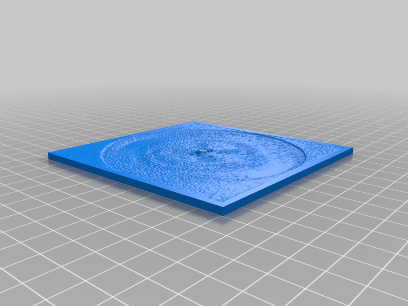
Original image file: https://en.wikipedia.org/wiki/File:Photo_51_x-ray_diffraction_image.jpg Converted to lithophane using: http://3dp.rocks/lithophane/ Rosalind Franklin's groundbreaking X-ray diffraction image of a paracristalline gel, known as Photo 51, was taken by her graduate student, Raymond Gosling, in May 1952 at King's College London. The photo, often misinterpreted as crystallized DNA, was part of a larger project led by Sir John Randall. Gosling had initially worked under Franklin's supervision but later returned to work directly under Wilkins' guidance after Franklin left King's and Randall requested that Gosling share all his data with Wilkins. Watson's collaborator, Maurice Wilkins, showed him the photo without Franklin's knowledge or consent. Watson and Crick used Photo 51, along with evidence from multiple other sources, to develop their groundbreaking chemical model of the DNA molecule in 1953. The model was first published together with manuscripts by Wilkins and his colleagues, as well as Gosling and Franklin, in the same issue of Nature. In recognition of their contributions, Watson, Crick, and Wilkins were awarded the Nobel Prize in Physiology or Medicine in 1962. Despite her significant role in the discovery, Rosalind Franklin did not receive the prize; she had passed away four years earlier, and the Nobel Committee does not make posthumous nominations. Gosling's work was also overlooked by the prize committee.
With this file you will be able to print Photo 51: x-ray diffraction pattern of DNA with your 3D printer. Click on the button and save the file on your computer to work, edit or customize your design. You can also find more 3D designs for printers on Photo 51: x-ray diffraction pattern of DNA.