Protein domains
thingiverse
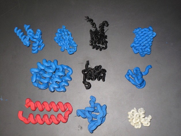
Proteins are polymers of amino acids. The primary sequence of amino acids conveys to the protein the ability to fold into particular structures in three-dimensional space. Thus a protein is a series of 3D domains strung together to give a protein a particular set of functions. For example, if death and EF hand domains are part of the same protein, the protein may trigger cell death with a rise in cellular calcium levels. New proteins are selected by evolutionary pressure when different domains are mixed and matched. I have uploaded a second version of CFP that is easier to remove the support material Instructions Students learn in many different ways. One important way to learn is tactile. Some people learn best by actually touching three-dimensional models. In biochemistry classes, students usually visualize protein structures using programs such as pymol, rasmol etc. For teaching protein structure and function, I have paired 3D printed models with pymol visualization in an iPad. There are over 40 different protein domains. The three dimensional structure of proteins and their domains is determined by x-ray crystallography or nuclear magnetic resonance (NMR). The 3D coordinates of each atom in a domain is determined and provided in a protein database (PDB) format. The PDB files for every proteins structure ever determined are freely available at PDB.org. Below is a brief description of ten different domains, followed by an explanation of how to take a PDB file and convert it to an STL file. A shout-out to PMOEWS whose work (thing:12283) inspired me and offered direction. SH2 domain #1BFJ Src-homology 2 (SH2) domains are modules of ~100 amino acids that bind to specific phospho tyrosine (pY) containing peptide motifs. Conventional SH2 domains have a conserved pocket that recognizes pY, and a more variable pocket that binds 3-6 residues C-terminal to the pY and confers specificity. SH3 domain #1NEB Src-homology 3 (SH3) domains bind to Pro-rich peptides that form a left-handed poly-Pro type II helix, with the minimal consensus Pro-X-X-Pro. Each Pro is usually preceeded by an aliphatic residue. Each in the aliphatic-Pro pair binds to a hydrophobic pocket on the SH3 domain. CARD domain #1CY5 Caspase Recruitment Domains (CARDs) are modules of 90–100 amino acids involved in cell death (apoptosis) signaling pathways. CARDs mediate the association of adaptor proteins and procaspases (death proteins) through heterodimerization of their respective CARDs, recruiting procaspases to upstream signaling complexes and allowing autoactivation. Death Domain #1DDF Death domains (DD) are 80–100 residues long motifs involved in cell death (apoptotic) signal transduction. Death domains serve as recruiting modules through their ability to heterodimerize with the death domains of distinct proteins, including adaptor proteins such as FADD. EF hand domain #2PMY The EF-hand motif contains approximately 40 amino acids (residues) and is involved in binding intracellular calcium. Binding of calcium to regulatory EF-hand domain—containing proteins induces a conformational change, which is transmitted to their target proteins, often catalyzing enzymatic reactions. Beta barrel (cyan fluorescent protein) #4AR7 This fluorescent protein is a variation of green fluorescent protein from a jellyfish and is the only domain that is a complete protein. The protein is routinely used to visualize a variety of biological processes. The beta barrel domain is a beta sheet wrapped around the fluorescent active site to provide structure. Ring domain #1CHC The RING finger is a specialized type of Zn finger consisting of 40–60 residues that binds two atoms of zinc, and is involved in mediating protein—protein interactions. Many zinc fingers bind nucleic acids. The presence of a RING finger domain is a characteristic of RING-class E3 ubiquitin protein ligases capable of transferring ubiquitin from an E2 enzyme to a substrate protein. WW domain #1EOM WW domains are small 38 to 40 amino acid residue modules that have been implicated in binding to Pro-rich sequences. Helix turn helix domain #3V1A The helix-turn helix is a DNA-binding domain. The two alpha helices are the reading or recognition helices, which bind in a groove in the DNA and recognize specific gene regulatory sequences in the DNA. This pairs well with thing:17343 by emmett! Ig domain #2CKN This particular domain is named for the first protein in which it was found, the immunoglobulin. An immunoglobulin is a antibody. Antibodies are generated by our immune system to recognize the specific size, shape and charge of pathogens. This domain is also found on the extracellular portion of many receptors including the interleukin-1 family of receptors. Convert pdb to stl file Select structure from pdb.org open VMD 1.9.1 (ks.uiuc.edu/Research/vmd/) Load pdb file →file “new molecule†“graphics†→ representations → drawing method “tube†Radius 1.5 Resolution 10 Display →axes → off File→ render → “enter file name†→ STL → render Open in replicator G Scale to appropriate size (many times very small and unseeable!) Upload to cloud.nettfabb.com to fix stl Slice and print!
With this file you will be able to print Protein domains with your 3D printer. Click on the button and save the file on your computer to work, edit or customize your design. You can also find more 3D designs for printers on Protein domains.